


Home / Training / Manuals / Atlas of breast cancer early detection / Cases
Atlas of breast cancer early detection
Go back to the list of case studies
.png) Click on the pictures to magnify and display the legends
Click on the pictures to magnify and display the legends
| Case number: | 180 |
| Age: | 67 |
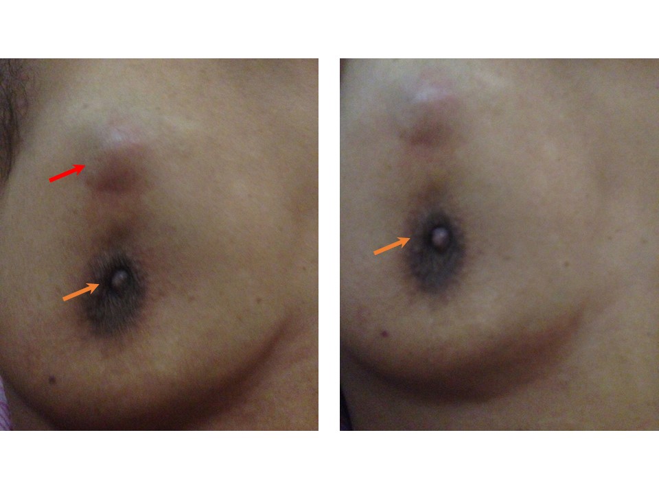
| Clinical presentation: | Postmenopausal woman with average risk of breast cancer presented with a right breast lump first noticed 12 years ago, which had progressively increased in size. On examination, a hard, non-tender lump was palpable in the right breast. It was fixed to the chest wall and skin and fixed to the breast parenchyma. The right nipple was retracted. The left breast was unremarkable. |
 |
Mammography:
 | 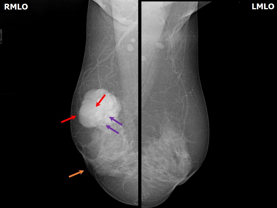 |
| Breast composition: | ACR category c (the breasts are heterogeneously dense, which may obscure small masses) | Mammography features: |
| ‣ Location of the lesion: | Right breast, upper quadrant at 12 oclock, middle and posterior thirds |
| ‣ Mass: | |
| • Number: | 1 |
| • Size: | 4.0 × 3.6 cm |
| • Shape: | Irregular |
| • Margins: | Microlobulated |
| • Density: | High |
| ‣ Calcifications: | |
| • Typically benign: | None |
| • Suspicious: | Suspicious calcifications |
| • Distribution: | Within mass |
| ‣ Architectural distortion: | None |
| ‣ Asymmetry: | None |
| ‣ Intramammary node: | None |
| ‣ Skin lesion: | None |
| ‣ Solitary dilated duct: | None |
| ‣ Associated features: | Nipple retraction, coarse heterogenous and pleomorphic calcifications within mass |
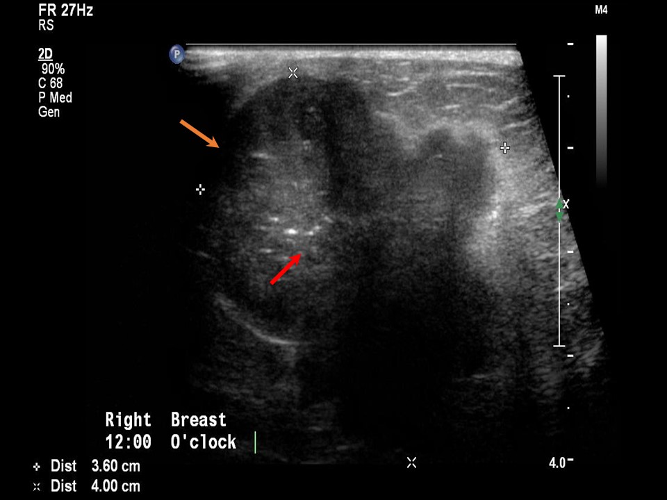
Ultrasound:
 |  |
 |
| Ultrasound features: Right breast, upper quadrants at 12 oclock, 2 cm from the nipple | |
| ‣ Mass | |
| • Location: | Right breast, upper quadrants at 12 oclock, 2 cm from the nipple |
| • Number: | 1 |
| • Size: | 4.0 × 3.6 cm |
| • Shape: | Irregular |
| • Orientation: | Not parallel |
| • Margins: | Angular |
| • Echo pattern: | Heteroechoic |
| • Posterior features: | No posterior features |
| ‣ Calcifications: | Pleomorphic and coarse heterogeneous calcifications in mass |
| ‣ Associated features: | Peripheral and central vascularity in mass |
| ‣ Special cases: | None |
BI-RADS:
BI-RADS Category: 5 (highly suggestive of malignancy)Further assessment:
Further assessment advised: Referral for core biopsyHistopathology:
Core needle biopsy
 |
| Histopathology features: | |
| ‣ Specimen type: | Core needle biopsy |
| ‣ Laterality: | Right |
| ‣ Macroscopy: | Three whitish cores 1.5 cm, 1.5 cm, and 1 cm long |
| ‣ Histological type: | Invasive breast carcinoma of no special type |
| ‣ Histological grade: | Grade 2 (3 + 2 + 1 = 6) |
| ‣ Mitosis: | 6 |
| ‣ Maximum invasive tumour size: | |
| ‣ Lymph node status: | |
| ‣ Peritumoural lymphovascular invasion: | |
| ‣ DCIS/EIC: | |
| ‣ Margins: | |
| ‣ Pathological stage: | |
| ‣ Biomarkers: | |
| ‣ Comments: |
MRM
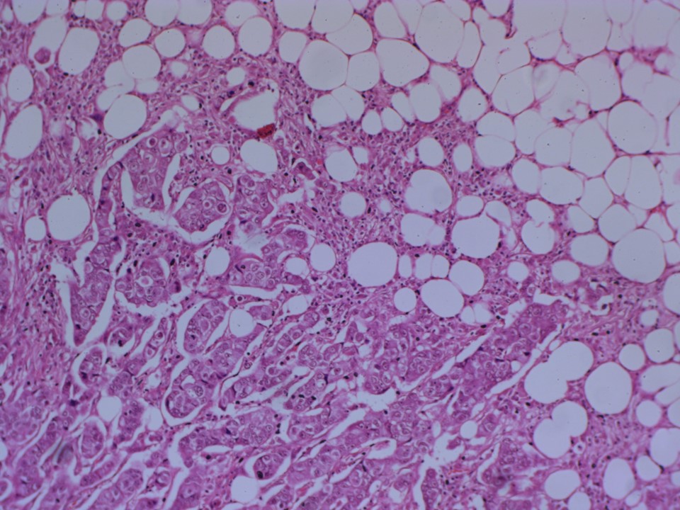 |  |
| Histopathology features: | |
| ‣ Specimen type: | MRM |
| ‣ Laterality: | Right |
| ‣ Macroscopy: | Right MRM specimen (30.0 × 18.0 × 4.0 cm) with overlying skin flap (13.5 × 7.5 cm). Nippleareolar area is unremarkable. On serial sectioning, a well-circumscribed grey white tumour (4.0 × 3.6 × 3.5 cm) is identified, located in the upper half superior to the areola. It is located 0.1 cm from the skin and 1.5 cm from the base. The attached axillary tail is 8 cm long. Ten lymph nodes were identified, the largest of which was 1.0 × 1.0 cm; the others ranged in size from 0.9 to 0.3 cm. Two apical nodes were received separately |
| ‣ Histological type: | Invasive breast carcinoma of no special type |
| ‣ Histological grade: | Grade 2 (3 + 3 + 1 = 7) |
| ‣ Mitosis: | 6 |
| ‣ Maximum invasive tumour size: | 4.0 cm |
| ‣ Lymph node status: | 0/12 |
| ‣ Peritumoural lymphovascular invasion: | Present |
| ‣ DCIS/EIC: | DCIS comedo-type high grade |
| ‣ Margins: | Free of tumour |
| ‣ Pathological stage: | pT2No |
| ‣ Biomarkers: | ER: Positive (100% of cells show nuclear staining with strong intensity. Allred score 8/8). PR: Positive (100% of cells show nuclear staining with moderate intensity. Allred score 7/8). HER2: Positive |
| ‣ Comments: | Tumour cells are seen in the dermis but do not involve the epidermis and there is no ulceration of the skin, so it does not qualify as pT4. Based on the size of the tumour it is pT2. |
Case summary:
| Postmenopausal woman presented with right breast lump with retracted nipple. Diagnosed as right breast carcinoma with coarse calcifications and fine pleomorphic calcifications within, right nipple retracted, BI-RADS 5 on imaging and as invasive breast carcinoma of no special type, pT2N0 on histopathology. |
Learning points:
|